 ALERT
ALERT ATTENTION ⚠️
In observance of a holiday, Agilent CrossLab/iLab Operations Software Support Help Desk will be closed during U.S. hours on Friday, July 3rd, 2026. We will resume regular U.S. support hours on Monday, July 6th, 2026. EU and APAC Support will remain open during this time. For urgent matters, please add "Urgent" to the ticket/email subject or press "1" when prompted to escalate a call on the iLab Support phone, and we will prioritize those requests first.
(hide this warning on this page)
|
|
Welcome to the Cell Sciences Imaging Facility at the Beckman Center The Cell Sciences Imaging Facility (CSIF) is a University service center that provides high resolution, state-of-the-art technologies for imaging and analyzing the molecular and structural organization of cells, tissue and bioengineered materials. The CSIF operates three sites at Stanford University: The CSIF at Beckman Center and the CSIF at Shriram Center, and the CSIF at Neuroscience (as NMS community lab). All locations are open to members of the Stanford community as well as to external academic and industry researchers (with approval). You are on the CSIF's Beckman iLabs Solutions site. The CSIF at Beckman houses both light and electron microscopy services. Light microscopy systems: Upright and inverted confocal laser scanning systems :Zeiss LSM880, AiryScan FAST; Leica SP8 gated-HyD, resonant, Lightning; upright LEICA Stellaris 8 DIVE (with long working distance immersion lens, tunable multiphoton laser), Zeiss Elyra7 lattice SIM, Nikon Eclipse Ti2-e motorized widefield and OMX SIM (TIRF) are accessible by users Widefield Nikon Eclipse for high resolution fluorescent and brightfield slide scanning with JOBS module. FESEM and TEM are available in our full service EM facility. Quick links to the microscopes scheduling calenders are below. The CSIF at Shriram provides access to and training on fluorescence and transmitted light microscopy including: laser scanning spectral confocal with AiryScan, multi-photon imaging (Leica Stellaris8 with Falcon), spinning disk confocal microscope with TIRF and photo-activation capabilities (Nikon SDC), FLIM (BH Simple Tau), Lattice lightsheet (Zeiss LLS7), Lightsheet (Bruker TrueLive) and widefield (epi-fluorescence, Zeiss AxioImager) microscopes. The CSIF at WuTsai make available instruments geared with neuroscience imaging in mind: laser scanning confocal microscopes with Airyscan2 (Zeiss 980+, Zeiss LSM980), LaVision lightsheet, Bruker/Prairie multiphoton rig, motorized Zeiss AxioImager, and Stellaromics Plexa Spatial Sequencer. Electron Microscopy Core (EMC) The CSIF-EMC is a full service lab providing access to and training on transmission (TEM) and scanning (SEM) electron technologies as well as sample preparation, immuno-histochemitry staining, room temperature electron tomography, serial-section SEM, EM imaging and documentation. Fluorescence Microscopy Core (FMC) The CSIF-FMC provides access to and training on high end fluorescent microscopes on an hourly fee basis. Current microscope technologies include super resolution (SIM, TIRF), laser scanning and spinning disk confocal, multi-photon, FLIM, TIRF and widefield deconvolution. Advance data processing workstations are housed here with: Imaris, Huygens, and Python-oriented open source Napari/Cellpose/CellProfiler/QuPath/Matlab -- GPU-supported open source options available in-person and remote-accessible. Multiplexing Core (MPC) Full-service codex multiplexing personnel provides service using Akoya Phenocycler-Fusion2.0. Workstations with advanced data processing available: Visiopharm, Fiji are GPU-supported options on the workstations here. Neuroscience Core (NSC) Serving the needs tailored toward neuroscience-imaging specific use, include animal-preparation room uniquely available at this location.
The CSIF (RRID:SCR_017787) is a member of and is supported by: The Beckman Center, Stanford Cancer Institute and BioX
|
Gordon Wang | CSIF Director
Office/Lab (B050): (650) 723-8818
Email: drwonder@stanford.edu
| Name | Role | Phone 1(650) | Location | |
| Gordon Wang | CSIF Director | 723-8818 | drwonder at stanford.edu | Beckman B062 |
| Jon Mulholland | CSIF Director Emeritus | jwm at stanford.edu | ||
| John Perrino | EM Lab Manager - TEM | 723-3462 | jperrino at stanford.edu | Beckman B001 |
| Kitty Lee |
CSIF Facility Manager, Light Microscopy Specialist - Confocals |
723-2449 | kamanl at stanford.edu | Beckman B050B |
| Yuanyuan Li | Senior Multi-omics Imaging Specialist - Phenocycler | 723-9925 | yuanyli at stanford.edu | Beckman B005 |
| Yili Zhu | Multi-omics Imaging Specialist - Phenocycler | 723-9925 | yilizhu at stanford.edu | Beckman B005 |
| David Lenzi | CSIF Shriram Manager | 724-8442 | dlenzi at stanford.edu |
Shriram B023 |
|
Hours |
|
|
Cell Sciences Imaging Facility Stanford University Medical Center Beckman Center B050, B005 (FMC) Beckman Center B001, B005, B050C, B051 (EMC, ATC) Stanford, CA 94305-5330 |
8 a.m. - 5 p.m., Monday to Friday 24/7 access for trained users
|
Reservation Calendars Quick Links
As with all Stanford Service Centers, credit must be given to Cell Sciences Imaging Facility for data that results in a publication. If the work done at Cell Sciences Imaging Facility produces data resulting in a figure in a publication, you are required to acknowledge Cell Sciences Imaging Facility in the publication. Further, if Cell Sciences Imaging Facility staff members provided significant experimental design, data interpretation, or other intellectual contribution (as evaluated by the PI), then it is expected that these individuals will be coauthors on the publication.
Proper Citation: Stanford University Cell Sciences Imaging Core Facility (RRID:SCR_017787)
Additionally almost all of the CSIF's microscopes were obtained via NIH instrumentation grants and therefore require proper acknowmedgement. Please see the required NIH citation information included on the microscope's description page or contact the CSIF director.
| Name | Role | Phone | Location | |
|---|---|---|---|---|
| Gordon Wang |
Director
|
650-723-8818
|
drwonder@stanford.edu
|
Beckman B062
|
| Jon Mulholland |
Director Emeritus
|
jwm at stanford.edu
|
||
| John Perrino |
EM Lab Manager
|
650-723-3462
|
perrino at stanford.edu
|
B001
|
| Kitty Lee |
Facility Manager
|
650-723-2449
|
kamanl at stanford.edu
|
B050
|
| David Lenzi |
Shriram Manager
|
650-724-8442
|
dlenzi at stanford.edu
|
Shriram B023
|
| Yuanyuan Li |
Senior Multi-omics Manager
|
650-723-9925
|
yuanyli@stanford.edu
|
B005
|
| Yili Zhu |
Multi-omics Scientist
|
650-723-9925
|
yilizhu@stanford.edu
|
B005
|
| Instrumentation, Services, & Training Price List |
| ► Array Tomography (ATC) Services (1) | |||
| Name | Description | Price | |
|---|---|---|---|
| Array Generation (per hour) | (view additional details) | Inquire | |
| ► CODEX services (1) | |||
| Name | Description | Price | |
| CODEX base run: Fresh Frozen | Inquire | ||
| ► EMC Equipment Fees (9) | |||
| Name | Description | Price | |
| Critical Point Dryer - CPD (per run) | (view additional details) | Inquire | |
| Cryo-ultramicrotome (per hour) | Inquire | ||
| Freeze substitution (chemicals included; per 2-3 day run) | Inquire | ||
| High Pressure Freezer (per half day; 4 hrs.) | (view additional details) | Inquire | |
| Specimen Processing Reagent Fee - FULL (per processing) | Inquire | ||
| Specimen Processing Reagent Fee - PARTIAL (per processing) | Inquire | ||
| Sputter Coater (per run) | (view additional details) | Inquire | |
| Ultramicrotome Leica UCT (per hour) | (view additional details) | Inquire | |
| Vacuum (Carbon) evaporator (per run) | (view additional details) | Inquire | |
| ► EMC Services (17) | |||
| Name | Description | Price | |
| EMC Technician Time (per hour) | (view additional details) | Inquire | |
| Grid Staining (per each additional grid) | (view additional details) | Inquire | |
| Grid Staining: Uranyl acetate and lead citrate contrast staining (up to 6 Grids) | (view additional details) | Inquire | |
| Immuno-gold Localization (Typical cost. Single antibody/grid on up to 8 grids, including controls) | (view additional details) | Inquire | |
| ImmunoEM Resin embedding | (view additional details) | Inquire | |
| Negative Staining (per additional grid) | (view additional details) | Inquire | |
| Negative Staining - includes reagents (up to 5 Grids) | (view additional details) | Inquire | |
| SEM Imaging (SEM time plus EM Microscopist time - per hour) | (view additional details) | Inquire | |
| SEM Sample OTO Prep for Serial Section Array (up to 6 samples) | (view additional details) | Inquire | |
| SEM Sample Prep - Specimen mounting and Sputter Coating (up to 6 samples) | (view additional details) | Inquire | |
| SEM Services: Sample Prep - Fixation and CPD Processing (up to 6 samples) | (view additional details) | Inquire | |
| TEM tomography: Single or Double tilt | (view additional details) | Inquire | |
| TEM Ultrastructural Services: Sample Prep - Cryo fixation, freeze substitution (2-6 samples; typical cost) | (view additional details) | Inquire | |
| TEM Ultrastructural Services: Sample Prep - Microwave-assisted chemical fixation (2-6 samples; typical cost) | (view additional details) | Inquire | |
| TEM Ultrastructural Services: Sample Prep - Standard chemical fixation and resin processing (2-6 samples; typical cost) | (view additional details) | Inquire | |
| Ultramicrotomy: block trimming (per sample-block, 4 grids) | (view additional details) | Inquire | |
| Ultramicrotomy: Previously trimmed by EM facility block (per sample-block, 4 grids) | (view additional details) | Inquire | |
| ► EMC Training (2) | |||
| Name | Description | Price | |
| EM TRAINING on SEM | Inquire | ||
| EM TRAINING on TEM | (view additional details) | Inquire | |
| ► Electron Microscopy Center (EMC) Microscopes (4) | |||
| Name | Description | Price | |
| Histology Scope: Leica DM2000 | Inquire | ||
| SEM: Hitachi S-3400 VP | Inquire | ||
| SEM: Zeiss Sigma FESEM | Inquire | ||
| TEM JEOL JEM-1400 | Inquire | ||
| ► FMC Microscopes (4) | |||
| Name | Description | Price | |
| LSM880 inverted confocal, Airyscan, GaAsP detector | Inquire | ||
| OMX Blaze SIM-STORM-Deconvolution | Inquire | ||
| SP8 inverted confocal, WLL, lightning, gSTED, FLIM | Inquire | ||
| Stellaris8 DIVE Confocal, multi-photon | Inquire | ||
| ► FMC Services and Training (3) | |||
| Name | Description | Price | |
| FMC Microscope Training & Usage Set-Up | (view additional details) | Inquire | |
| FMC Technical Assistance / Consulting (per hour) | (view additional details) | Inquire | |
| Work Station 1,2,3,4 Usage | (view additional details) | Inquire | |
| ► FMC supplies (8) | |||
| Name | Description | Price | |
| Bioptech chamber | (view additional details) | Inquire | |
| Fluoro Dish 23mm (small) | (view additional details) | Inquire | |
| Fluoro Dish 40mm (large) | (view additional details) | Inquire | |
| High performance #1.5 cover glass | (view additional details) | Inquire | |
| Materials | (view additional details) | Inquire | |
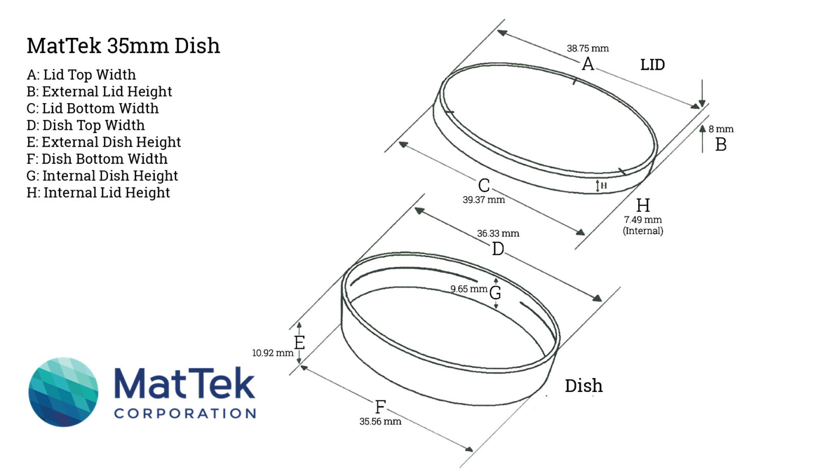
| Mattek Chamber | (view additional details) | Inquire | |
| Nunc Chamber | (view additional details) | Inquire | |
| POC-R coverslip | (view additional details) | Inquire | |
| ► Array Tomography (ATC) Equipment (1) | |||||||||||||||||||||||||||||||||
|
|||||||||||||||||||||||||||||||||
| ► Electron Microscope Core (EMC): Equipment (10) | |||||||||||||||||||||||||||||||||
|
|||||||||||||||||||||||||||||||||
| ► Electron Microscope Core (EMC): Microscopes (4) | |||||||||||||||||||||||||||||||||
|
|||||||||||||||||||||||||||||||||
| ► Fluorescence Microscope Core (FMC): Microscopes/Equipment (10) | |||||||||||||||||||||||||||||||||
|
|||||||||||||||||||||||||||||||||

